Dental X-rays are very safe. The radiation exposure from a standard set of dental X-rays is extremely low. Comparable to the amount of background radiation a person naturally receives in a single day. The doctors at BayView Dental Arts, including prosthodontist and founder Dr. Edward A. Scherder, DMD, JD, prioritize patient safety at every step, using the latest digital sensor technology to minimize radiation exposure.
In this blog, we will discuss how dental X-ray radiation works, the most common types of dental radiographs, how digital sensors protect patients, and how the team at BayView Dental Arts keeps Naples patients informed and safe.
What Is Radiation, and Why Is It Used in Dentistry?
Radiation is energy that travels in waves or particles. In dentistry, a small, controlled beam of X-ray energy passes through the mouth to capture images of teeth, bone, and surrounding structures that are invisible during a standard visual exam. According to the American Dental Association, how often X-rays should be taken depends on individual factors, including current oral health, age, risk for disease, and any signs or symptoms of concern, meaning there is no one-size-fits-all approach.
Key reasons dental X-rays are recommended include:
- Detecting cavities between teeth: Which are not visible to the naked eye during a routine exam.
- Evaluating bone loss: Associated with gum disease or tooth loss.
- Assessing tooth roots and surrounding bone: Before root canals, extractions, or implant placement.
- Monitoring growth and development: Particularly in younger patients with emerging permanent teeth.
- Planning restorative or implant treatment: To ensure precision and optimal long-term outcomes.
What Are the Different Types of Dental Radiographs?
Not all dental X-rays are the same. Different clinical situations call for different imaging tools, each with its own purpose and exposure level. The different types include:
- Periapical X-rays (PAs): Capture the full length of one or a few individual teeth and the surrounding bone. Used to identify infection, root fractures, or bone changes around specific teeth.
- Full Mouth Series (FMX): A complete set of periapical and bitewing images that gives the dentist a full picture of every tooth and the supporting bone structure. Commonly taken for new patients or as part of a periodic comprehensive evaluation.
- Cone Beam CT (CBCT): A three-dimensional imaging tool that produces detailed scans of the teeth, jaw, and surrounding facial structures. Used primarily for implant planning, complex extractions, and evaluation of bone volume. The ADA recommends CBCT only when lower-exposure options cannot provide the necessary diagnostic information.
The doctors at BayView Dental Arts select the appropriate radiograph type based on each patient's individual clinical needs.
How Much Radiation Do Dental X-rays Actually Emit?
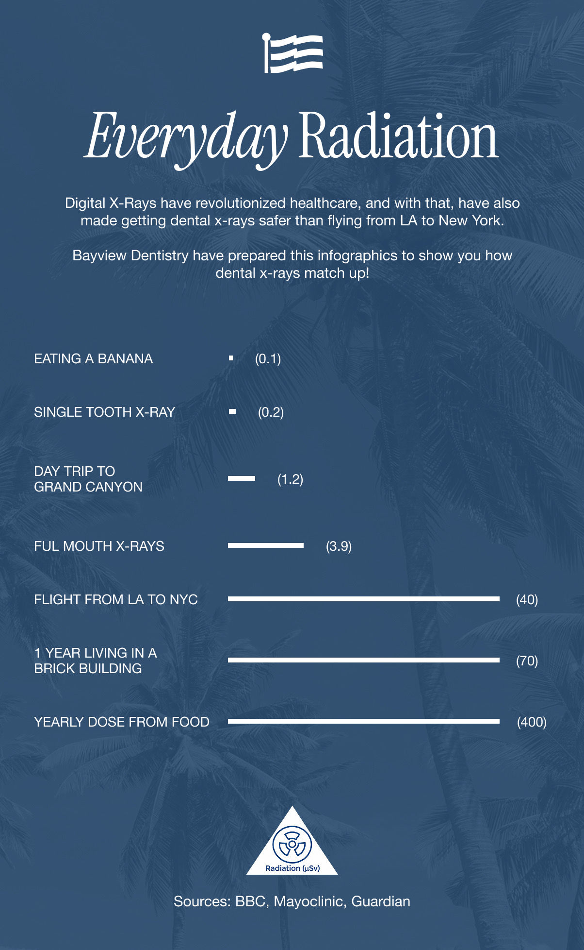
The dose is remarkably small. The American Dental Association notes that the average American receives approximately 6.2 millisieverts (mSv) of radiation per year from all sources combined, natural and man-made. Dental imaging accounts for less than 1% of the estimated collective annual effective dose received from medical imaging overall.
To put common dental X-rays in perspective:
- A single periapical (PA) X-ray: Delivers a fraction of the radiation from one day of natural background exposure.
- A full mouth series (FMX): Exposes a patient to approximately 0.1 mSv. Less than a cross-country flight.
- A CBCT scan: Involves more radiation than 2D imaging, but remains far below the exposure of a standard medical CT scan. The ADA recommends its use only when the clinical benefit clearly outweighs the additional exposure.
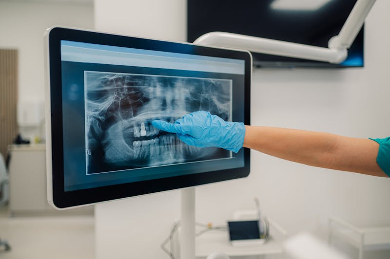
How Do Digital Sensors Reduce Radiation Exposure?
This is where BayView Dental Arts sets itself apart from practices that still rely on older technology. The practice uses the latest digital sensors for all radiographic imaging — a meaningful upgrade from traditional film that reduces radiation exposure significantly while producing sharper, more detailed images.
Specific advantages of digital sensors include:
- Up to 80% less radiation than conventional film: Making every X-ray, from a single PA to a full mouth series, as low-dose as clinically possible.
- Immediate, high-resolution image capture: Allowing the care team to review and discuss findings with patients in real time, without repeat exposures.
- No chemical processing required: A more environmentally responsible approach than film-based imaging.
- Enhanced diagnostic clarity: With the ability to zoom, adjust contrast, and annotate images for a more precise assessment.
Have Questions About Dental X-rays? The Doctors at BayView Dental Arts Are Here to Help.
Dental X-rays are among the most studied and tightly regulated tools in modern dentistry, and when delivered with today's digital sensor technology, the radiation exposure is minimal by any meaningful standard.
Whether you have questions about a specific type of radiograph or are simply due for your next exam, the team at BayView Dental Arts will walk you through every step with boutique-level care Naples patients have trusted for years. Call (239) 360-5944 or visit bayviewdentalarts.com to schedule your consultation today.